Figure 5.
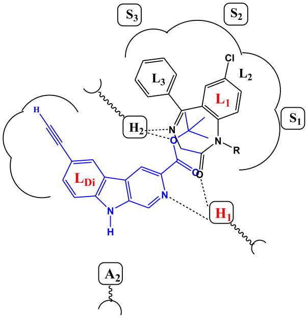
Figure 5a. Overlap of diazepam and βCCt in the pharmacophore/receptor model.
The structure of WYS8 and diazepam in a simple representation of the pharmacophore model. WYS8 (7) (blue line) and diazepam (black line) fitted to the inclusive pharmacophore model for the BzR. Sites H1 and H2 represent hydrogen bond donor sites on the receptor protein complex, while A2 represents a hydrogen bond acceptor site necessary for potent inverse activity in vivo. L1, L2, L3 and LDi are four lipophilic regions in the binding pharmacophore. Descriptors S1, S2, and S3 are regions of negative steric repulsion.
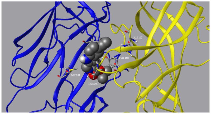
Figure 5b. WYS8 docked in the BzR site of the α1 subtype GABAA receptor. The α1 and γ2 subunits are rendered in yellow and blue, respectively.


