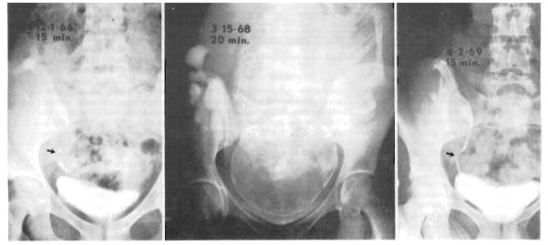
Fig. 1.
Intravenous pyelograms. Left, Seventeen months after renal homotransplantation and seven months before pregnancy. Note ureteroureterostomy (arrow). Center, During labor. Pyelogram was obtained in the course of pelvimetry. Right, Thirteen months post partum. Appearance is now similar to before pregnancy (left).

