Figure 6.
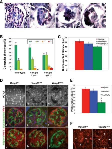
Disruption of Vangl2 causes glomerular maturation defects. All data in this figure are from E18.5 kidneys. Glomerular tuft morphology was assessed according to the following categories: a, more than two capillary lumens per tuft; b, one or two capillary lumens; c, no capillary lumens; d, abnormal and disrupted glomerular tuft. (A) Representative glomeruli in each category. (B) Proportions of glomeruli in the three genotypes (n = 3–4 kidneys in each set). The commonest phenotype of wild-type and heterozygous glomeruli was category a. Vangl2Lp/Lp glomeruli showed a shift of phenotypes such that glomerular types a and b were equally common and they contained a small subset of category d never seen in wild-type kidneys. Vangl2Lp/Lp glomerular tufts had a significantly reduced glomerular diameter (C). (Da–c) Proliferation of glomerular cells as assessed by immunodetection of PCNA (white nuclei). A reduction in the frequency of proliferation in homozygous organs was confirmed upon counting proportions of proliferating cells in the glomerular tuft (E). (Dd–f). Synaptopodin (green) and WT-1 (red) co-staining showed similar appearances in the three genotypes when mature glomeruli were assessed. (Dg–i) Nephrin (green) immunostaining showed similar appearances in the three genotypes when mature glomeruli were assessed. (F) Phalloidin staining highlighted the typical crenellated pattern of f-actin in Vangl2+/+ glomerulus (arrowed on the left frame) which appeared less apparent in a Vangl2Lp/Lp glomerulus (arrowed on the right frame). Counts were performed on at least 20 glomeruli from at least three to four separate kidneys in each genotype. *P < 0.05 compared with wild-type; +P < 0.05 compared with heterozygotes. Scale bars = 15 μm.

