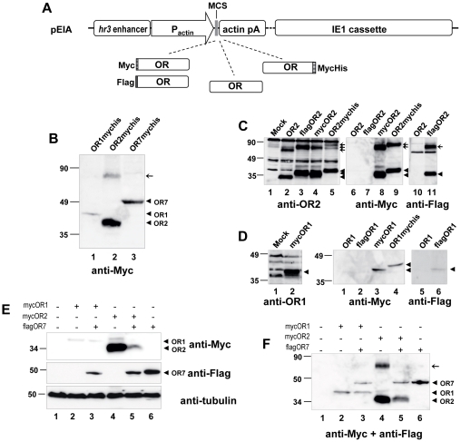
Figure 1. Expression of A. gambiae OR1, OR2 and OR7 in insect cells.
(A) Schematic representation of the basic backbone vector (pEIA) used for the heterologous expression of various forms (tagged and untagged) of ORs in lepidopteran cells. hr3 enhancer, baculoviral (BmNPV) homologous region 3 enhancer sequence; pActin, Bombyx mori A3 cytoplasmic actin promoter; MCS, multiple cloning site; actin pA, 3′untranslated region of B. mori actin gene containing polyadenylation signals; IE1 cassette, baculoviral (BmNPV) DNA fragment containing the ie-1 transactivator gene under the control of its native viral promoter; OR; A. gambiae odorant receptor ORF; Myc, Flag and MycHis, epitope tags. (B) Detection of heterologous expression of C-terminally MycHis-tagged ORs in transfected Hi5 cells using Myc monoclonal antibody. (C) Detailed western blot analysis of OR2. Hi5 cells were transfected with plasmids expressing different versions of OR2, and lysates were analyzed using a specific polyclonal antibody against OR2 (left panel, lanes 1–5) or monoclonal antibodies against the Myc (middle panel, lanes 6–9) or the Flag epitope (right panel, lanes 10–11). Arrowheads and arrows indicate major bands corresponding to monomers and putative dimers, respectively. (D) Detailed western blot analysis of OR1. Hi5 cells were transfected with plasmids expressing different versions of OR1, and lysates were analyzed using monoclonal antibodies against the Myc (middle panel, lanes 1–4) or the Flag epitope (right panel, lanes 5–6). In the left panel immunoreactivity of the specific polyclonal antibody against OR1 is shown, with lysates from cells expressing mycOR1 after treatment with the proteasome inhibitor MG132. Molecular weight markers are shown on the left. (E) and (F) Effect of coexpression of OR7 on the expression levels of OR1 and OR2. Hi5 cells were transfected with constant amounts (45% of total DNA) of Myc-tagged OR1 or OR2, along with equal amounts of Flag-tagged OR7 or empty vector (pEIA), and pEIA-GFP (10% of total DNA, for evaluation of the efficiency of transfection). Whole cell lysates (E) and membrane fractions (F) were analysed by SDS-PAGE and western blot. Detection of OR1, OR2 and OR7 was done using the anti-Myc and anti-Flag antibodies either consecutively (in E) or simultaneously (in F). To control for loading, the whole lysate fractions were also probed with an anti-tubulin antibody.

