Fig. 1.
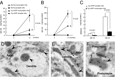
Increased accumulation of Aβ in synaptic mitochondria (mito) of Tg mAPP mice. (A and B) Age-related mitochondrial Aβ1-40 and Aβ1-42 accumulation in synaptic and nonsynaptic mitochondria from non-Tg and APP mice at 4 and 12 mo of age (n = 4–6 mice per group). *P < 0.05 vs. 4-mo-old Tg mAPP synaptic mitochondria; #P < 0.05 vs. 4-mo-old Tg mAPP nonsynaptic mitochondria. (C) Comparison of Aβ levels in synaptic mitochondria with those in nonsynaptic mitochondria from Tg mAPP mice at the age of 4 mo. (D–F) Immunogold EM images with a specific Aβ1-42 antibody, followed by a gold-conjugated antibody (18 nm), demonstrating the presence of intramitochondrial Aβ accumulation (black particles) in 12-mo-old Tg mAPP mice. Arrows denote mitochondria (M), and the asterisk denotes a synapse. (Scale bars = 200 nm.)

