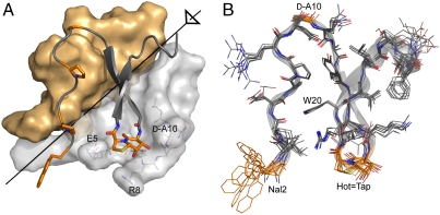
Fig. 4.
(A) Crystal structure of the trimeric FV-1. Two subunits are shown as surface representations (gray/yellow), one including three side chains (light gray) that embrace the Hot═Tap mimic of the third subunit (peptide backbone with selected side chains shown). The threefold axis is indicated in black. (B) Superposition of the backbones of all six different monomeric subunits in the asymmetric unit of the crystal structure of FV-1. Selected side chains are highlighted in orange.

