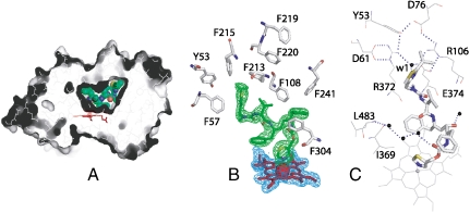
Fig. 6.
Crystal structure of the CYP3A4-ritonavir complex. (A), The active site cavity of ritonavir-bound CYP3A4. Ritonavir is green and in CPK representation; the heme is red. (B), Aromatic residues surrounding ritonavir. 2Fo-Fc (blue) and Fo-Fc (green) electron density maps around the heme and ritonavir are contoured at 1 and 3 σ, respectively. (C), An umbrella-like charge-charge/H-bonding network connected to the isopropyl-thiazole moiety of ritonavir via a highly ordered water molecule (w1).

