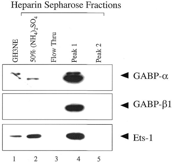
Figure 3.

Analysis of heparin–Sepharose purified GH3NEs by western blotting. Equal amounts (25 µg) of each heparin–Sepharose fraction, along with 25 µg of GH3NE for the GABP westerns, and 50 µg of GH3NE for the Ets-1 and Ets-2 westerns, were resolved on 10% SDS–polyacrylamide gels and transferred to Immobilon-P membranes. Blots were probed with antibodies against GABPα, GABPβ or Ets-1, where indicated. Protein was detected using ECL, as described in the Materials and Methods.
