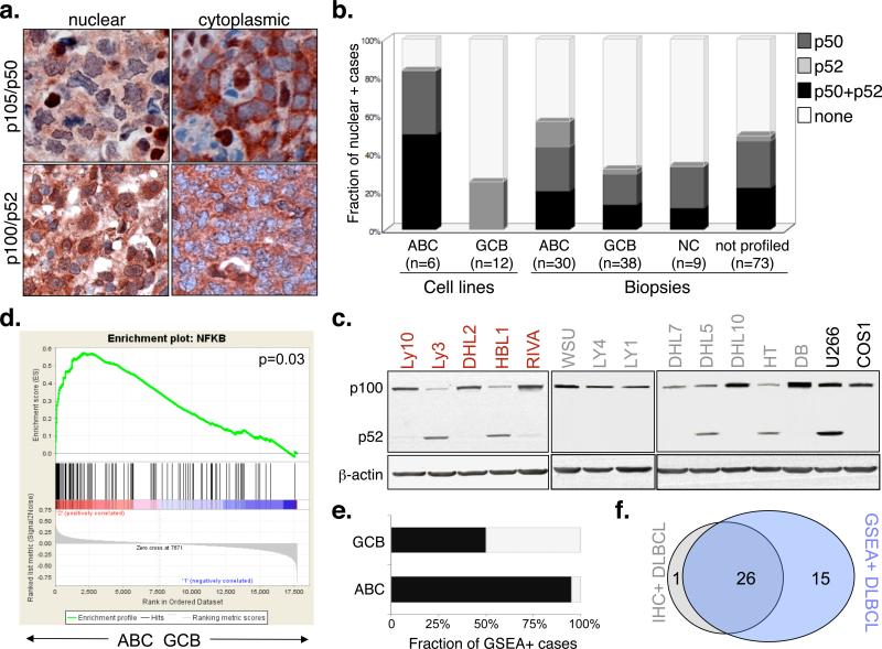
Figure 1. The NF-kB pathway is active in ABC-DLBCL and in a smaller fraction of GCB-DLBCL.
a) Immunohistochemical staining of DLBCL biopsies with anti-NFKB1(p105/p50)(top) and anti-NFKB2(p100/p52)(bottom) antibodies. Nuclear localization of NF-kB denotes active signaling, as opposed to the inactive, cytoplasmic complex (200X). b) Prevalence of cases displaying constitutive NF-kB activation in DLBCL subgroups. Color codes indicate nuclear p50, p52 or both. c) Western blot analysis of DLBCL cell lines for processing of p100 to p52 (red, ABC-DLBCL; grey, GCB-DLBCL). The multiple myeloma cell line U266 and the epithelial cell line COS are used as positive and negative controls, respectively. Actin, loading control. d) GSEA enrichment score and distribution of NF-kB target genes along the rank of transcripts differentially expressed in ABC- vs GCB-DLBCL. e) Percentage of samples showing a transcriptional NF-kB signature by GSEA. f) Venn diagram illustrating the overlap between immunohistochemistry-defined and GSEA-defined NF-kB positive cases.

