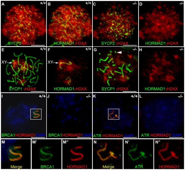
Figure 7. HORMAD1 disrupts γH2AX, BRCA1, and ATR localization to the XY chromosomes.
Chromosome spread assay was performed on wild-type and Hormad1−/− leptotene (A-D), zygotene (E-H) and pachytene (I-L) spermatocytes to determine effects of HORMAD1 deficiency on γH2AX, BRCA1 and ATR localization to the sex chromosomes. (A-D) Immunofluorescence assay with anti-SYCP2 (Green) or anti-HORMAD1 (Green) and anti-γH2AX (Red). (E-H) Immunofluorescence assay with anti-SYCP1 (Green) or anti-HORMAD1 (Green) and anti-γH2AX (Red) antibody. (C and D) At the leptotene stage, γH2AX and SYCP2 localization to chromatin can be detected in Hormad1−/− spermatocytes, however, γH2AX is substantially reduced. (F and H) γH2AX staining localizes preferentially to the sex chromosome in the wild-type pachytene stage spermatocytes but no preferential localization to the sex chromosomes was observed in the Hormad1−/− spermatocytes, which is not surprising since sex body does not form in Hormad1 mutants. (G) Arrow head indicates truncated axial fiber. (I, M-M”) BRCA1 and HORMAD1 co-localize to the sex chromosomes, but Hormad1 deficiency disrupts BRCA1 localization (J). Similarly, ATR co-localizes with HORMAD1 to the XY chromosomes (K, N-N”), and Hormad1 deficiency disrupts ATR localization (L). These results indicate that HORMAD1 is upstream of the currently known critical components of the meiotic sex chromosome inactivation complex, γH2AX, BRCA1 and ATR. DNA was stained with DAPI (Blue), and arrows indicate sex chromosomes. Scale bars: 10 µm.

