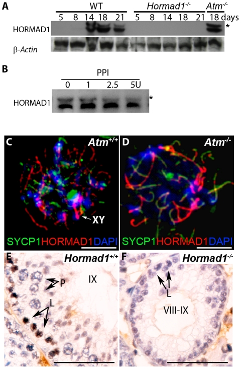
Figure 9. HORMAD1 phosphorylation is unaffected by Atm deficiency while Hormad1 deficiency disrupts ATM autophosphorylation.
(A) Western blots analyses with anti-HORMAD1 specific antibodies show that HORMAD1 protein and its phosphorylated form (*) appear circa post-natal day 14. Atm1 deficient testes (Atm− /−) express both forms of HORMAD1 and HORMAD1 is therefore unlikely to be ATM1 substrate. (B) The presumed phosphorylated form of HORMAD1 (higher molecular weight band indicated by asterisk) decreases in intensity after protein phosphatase I (PPI) treatment. (C and D) Chromosome spread assay in wild-type (Atm+/+), and Atm deficient spermatocytes (Atm−/−). HORMAD1 preferentially localizes to unsynapsed regions of chromosome axes (C), and HORMAD1 antibody stains unsynapsed chromosome axes in Atm−/− spermatocytes (D) more intensely than in the wild-type spermatocytes. Scale bars: 10 µm. (E and F) Immunohistochemistry analysis in 6 week old wild-type and Hormad1−/− testes with anti-phospho ATM-S1981 antibody. Phospho-ATM S1981 was detected in wild-type zygotene, but not detected in Hormad1−/− zygotene spermatocytes. Scale bars: 50 µm.

