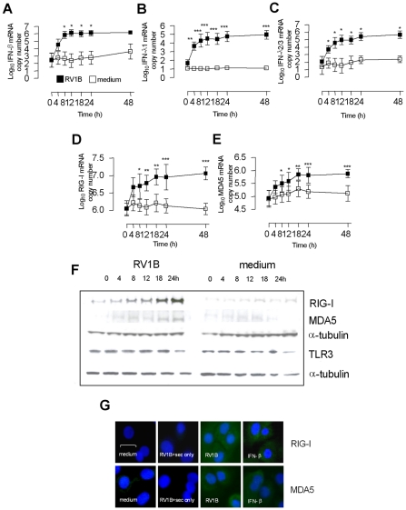
Figure 1. Kinetics of RV1B induced IFN-β, IFN-λs, RIG-I and MDA5 mRNA and protein expression in HBECs.
HBECs were infected with RV1B or treated with medium and RNA and protein analysed over time. RV1B induced IFN-β (A), IFN-λ1 (B) and IFN-λ2/3 mRNA (C) in a time dependent manner, visible by 4 h, and peaking at 12–48 h. RV1B infection also induced RIG-I (D) and MDA5 mRNA (E), in a time dependent manner, peaking at 18–48 h. RV1B infection also induced RIG-I and MDA5 protein (F) in a similar time dependent manner, visible by 8 h post infection by western blotting. Medium treated cells exhibited little or no RIG-I or MDA5 protein during the timecourse. TLR3 protein levels were present in medium treated cells, and did not change during the course of RV1B infection. Immunofluorescence identified both cytoplasmic RIG-I and MDA5 to be induced after RV1B or IFN-β treatment at 24 h, compared to medium treated cells, or cells stained with secondary antibody only. Horizontal line indicates 20 µm scale. Staining for both helicases was observed within the cytoplasm (in green, G). *p<0.05, **p<0.01, ***p<0.001, RV1B infected versus medium treated cells. mRNA data was generated from 6 independent experiments utilizing 3 independent HBEC donors, 2 experiments per donor.

