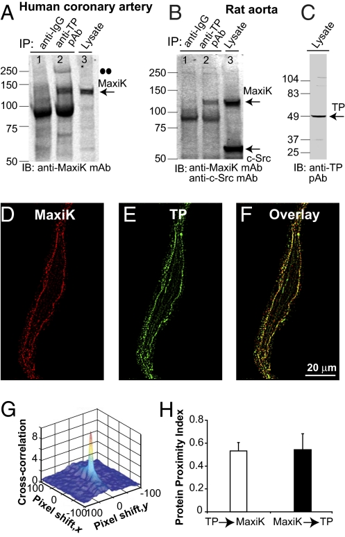
Fig. 5.
Vascular TP and MaxiK form a protein complex. (A and B) TP coimmunoprecipitates MaxiK in lysates of human coronary arteries and rat aorta (lane 2). Anti-rabbit IgG does not coimmunoprecipitate MaxiK (lane 1). (A and B, lane 3, and C) Input coronary and aorta lysates (40 and 50 μg protein, respectively). Double dots mark putative MaxiK dimer. (D and E) Freshly dissociated human coronary arterial myocyte double-labeled with anti-MaxiK monoclonal (red) and anti-TP polyclonal (green) antibodies. (F) Overlay (yellow). Images at 0.0288 μm/pixel after median filter background reduction. (G) TP and MaxiK 3D cross-correlation plot as a function of pixel shift. (H) Mean values of protein proximity index.

