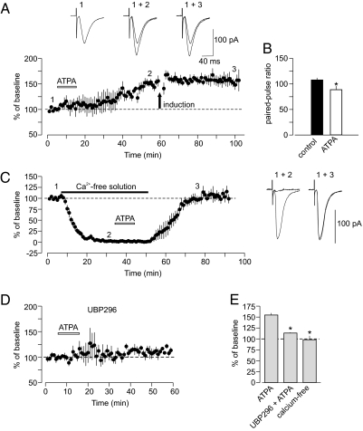
Fig. 2.
ATPA-induced potentiation and pre-LTP in thalamic input are mechanistically similar. (A) The GluR5-specific agonist ATPA (1 μM) in the external solution potentiated the EPSC in thalamic input (n = 4; P < 0.01 vs. baseline). Potentiation induced by ATPA has occluded LTP in response to electrical stimulation (delivered at arrow). (Insets) Averaged thalamo-amygdala EPSCs recorded under control conditions (1), during ATPA-induced potentiation (2), and after the delivery of LTP protocol (3). (B) Paired-pulse facilitation (PPF; 50-ms interpulse interval) was decreased during ATPA-induced potentiation (n = 4, P = 0.017). (C) Potentiation was prevented when ATPA (1 μM) was applied in Ca2+-free external solution (n = 6, P = 0.75 for post-ATPA vs. baseline). Traces (to the right) are averaged EPSCs recorded at different time points (shown as 1, 2, and 3) during the experiment. (D) UBP296 (1 μM) blocked potentiation of thalamo-amygdala EPSCs by ATPA (n = 3). (E) Summary of experiments with ATPA (ATPA, n = 4; UBP296 + ATPA, n = 3, P = 0.014 vs. ATPA alone; ATPA in Ca2+-free solution, n = 6, P = 0.01 vs. ATPA alone). Error bars indicate SEM.

