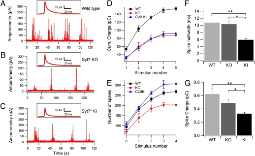
Fig. 1.
Conventional amperometry reveals a decrease in Ca2+-triggered exocytosis in Syt7 KO and Syt7* KI chromaffin cells. (A–C) Representative traces of bursts of amperometric spikes in WT (A), Syt7 KO (B), and Syt7* KI chromaffin cells (C). Spikes were triggered by consecutive puffs of 70 mM K+ from an adjacent pipette. (D) Integral of amperometric responses during successive stimulations. Notice that Syt7 KO and KI produced a smaller response for all stimuli in the series. Catecholamine release is decreased approximately twofold in Syt7 KO and Syt7* KI chromaffin cells as compared with WT cells (P = 0.036 and P = 0.032, respectively). (E) Number of amperometric spikes during successive stimulations. Syt7* KI cells produced a larger number of spikes per stimulation than the Syt-7 KO (P = 0.0024) and the WT (P = 0.021) cells. (F and G) Kinetic parameters of amperometric spikes in WT, Syt7 KO, and Syt7* KI chromaffin cells. (F) Spike half-widths, measured per individual amperometric spikes, were similar in WT and Syt7 KO cells but were significantly smaller in the Syt7* KI than in WT cells (**P = 0.00048) or the Syt7 KO (*P = 0.00016). (G) Spike charge was significantly different in WT and Syt7* KI cells (**P = 0.00005) and in Syt7 KO and Syt7* KI cells (*P = 0.03).

