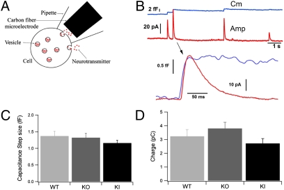
Fig. 3.
Patch amperometry of single full-fusion exocytotic events in Syt7 mutants. (A) Patch amperometry configuration. On-cell patch clamping is achieved with a pipette containing a CFE. The cell membrane capacitance can be measured with a resolution that detects fusion of a single chromaffin granule with the membrane patch. Release of catecholamines into the patch pipette by spontaneous exocytotic events is measured electrochemically with the CFE. Catecholamine release occurring over the rest of the cell surface is not detected. (B) (Upper) Representative traces (example depicts a WT cell) showing simultaneous measurements of step increase in capacitance (blue trace) and of amperometric spike (red trace). (Lower) In the expanded exocytotic event, notice the delay between the onset of the capacitance and amperometric increase. (C) The size of the capacitance steps induced by exocytosis of individual chromaffin granules was similar for WT, Syt7 KO, and Syt7* KI cells (P > 0.05). (D) The charge in individual amperometric spikes associated with full-fusion events as detected by capacitance was similar for WT (3.2 ± 0.5 pC) and Syt7 KO cells (3.8 ± 0.4 pC, P > 0.05) but decreased slightly in Syt7* KI cells (2.7 ± 0.3 pC) compared with Syt7 KO cells, consistent with the reduced charge measured in Syt7* KI cells by conventional extracellular amperometry.

