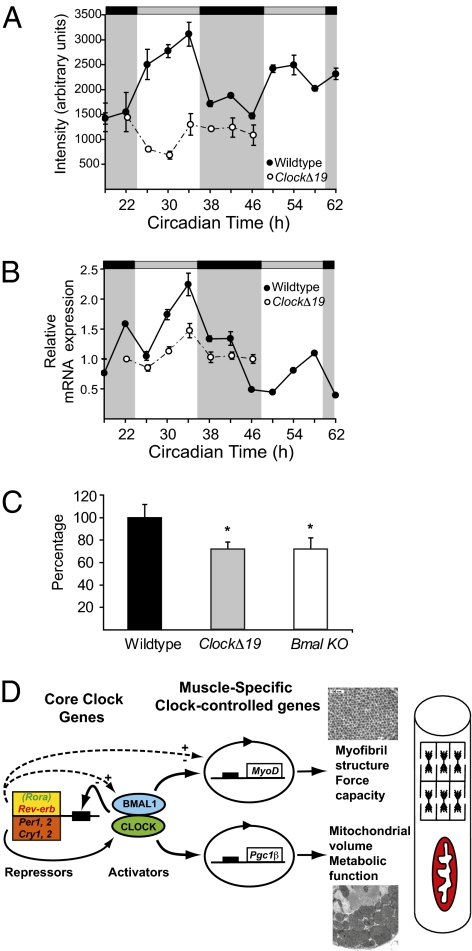
Fig. 4.
Altered expression of Pgc-1 coactivators in ClockΔ19 and Bmal1−/− mice. (A) Array data of Pgc-1β mRNA expression in skeletal muscle of wild-type mice (●) and ClockΔ19 mice (○); the light and dark stripes refer to the presumptive light and dark phases for the mice (7). (B) Quantitative PCR results for expression of Pgc-1β in wild-type muscle (●) and ClockΔ19 muscle (○). (C) Histogram of the mean expression level of PGC1α mRNA in muscle of wild-type, ClockΔ19, and Bmal1−/− mice as determined by quantitative PCR. A significant difference (P < 0.05) is denoted by an asterisk. (D) Proposed model of CLOCK:BMAL1 regulation of muscle phenotype and function via targeting of MyoD and Pgc-1 expression. Solid lines indicate known molecular links among components of the molecular clock, and dashed lines suggest potential links.

