Fig. 1.
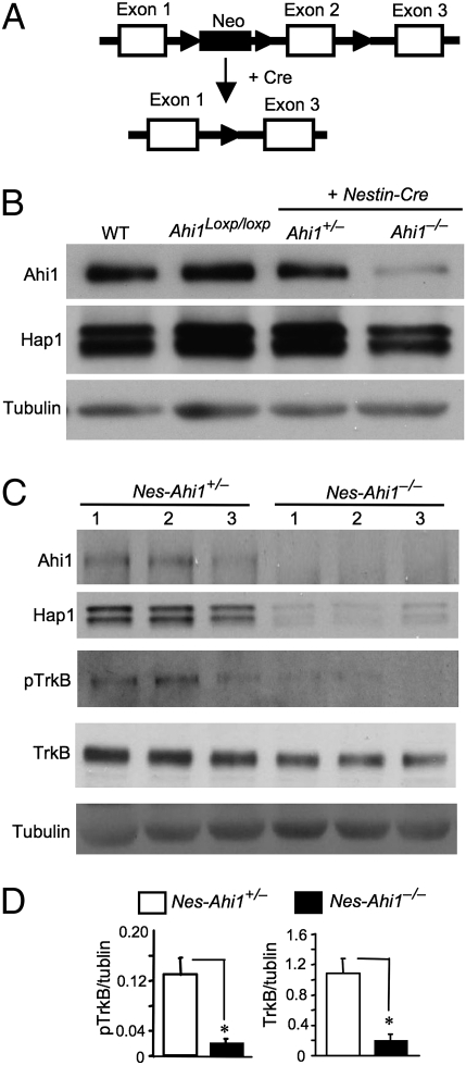
Ahi1 deficiency diminishes Hap1 and TrkB. (A) Generation of conditional Ahi1 knockout mice. Ahi1 exon 2 was removed by the Cre–loxP system to suppress Ahi1 expression. (B) Western blot analysis of Ahi1 and Hap1 expression in the hypothalamic tissue from wild-type (WT), floxed Ahi1 (Ahi1loxp/loxp), heterozygous (nes-Ahi1+/−), and homozygous conditional knockout (nes-Ahi1−/−) mice. (C) Total TrkB and pTrkB in the hypothalamus of control (nes-Ahi1+/−) and Ahi1-deficient (nes-Ahi1−/−) mice were examined by Western blotting. The blots were also probed with antibodies to Ahi1, Hap1, and tubulin. (D) Densitometric analysis of the ratios of TrkB or pTrkB to tubulin showing a decrease of total TrkB and pTrkB in Ahi1-deficient (nes-Ahi1−/−) mice (n = 3 each group, *P < 0.05).

