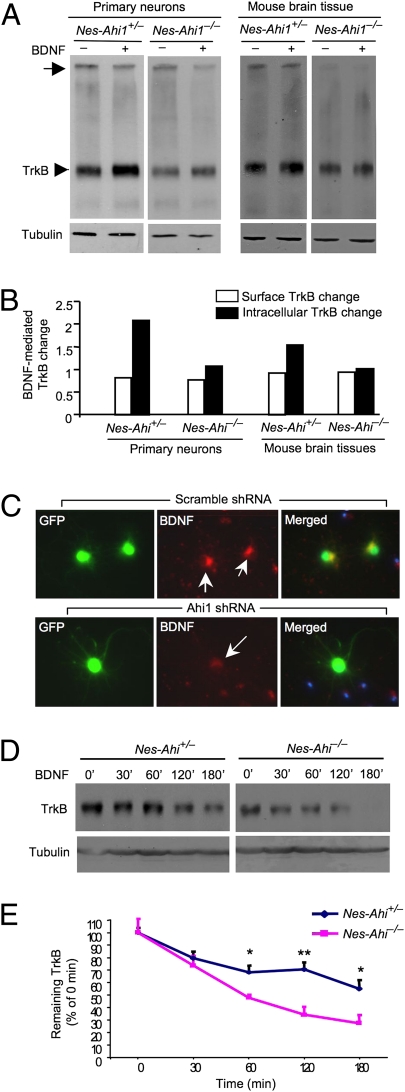
Fig. 4.
Ahi1 deficiency reduces the internalized TrkB and facilitates TrkB degradation. (A) Western blotting and BS3 crosslinking of membrane-bound receptors showing a reduction in membrane TrkB (arrow) and intracellular TrkB (arrowhead) in cultured primary brainstem cells and brainstem sections from nes-Ahi1−/− mice. BDNF (100 ng/mL for 1 h) was used to trigger TrkB internalization. (B) Quantifying BDNF-mediated changes of TrkB relative to that before BDNF treatment. (C) Decreased internalization of BDNF in cultured mouse brainstem cells treated with adenoviral Ahi1 shRNA. Arrows indicate neurons that coexpress adenoviral Ahi1 shRNA and GFP. (D) Cultured brainstem cells from control (nes-Ahi1+/−) mice and Ahi1-deficient (nes-Ahi1−/−) mice were treated with BDNF to induce TrkB internalization. TrkB Western blotting was then performed to assess the levels of TrkB at different times. (E) Quantitative analysis of the degradation of TrkB in brainstem slices of nes-Ahi1+/− and nes-Ahi1−/− mice. The ratios of TrkB to tubulin were measured via densitometry. The relative TrkB expression level at different times is expressed as percentage of the TrkB/tubulin ratio at 0 min before BDNF-induced internalization. The data were obtained from three independent experiments. *P < 0.05.

