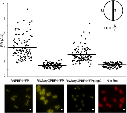
Fig. 2.
Septal localization of PBP4 is lost in a tagO null mutant. Microscopy images and quantification of septum versus lateral membrane fluorescence (fluorescence ratio, FR) of PBP4–YFP in a wild-type background (RNPBP4YFP), a ΔtagO background (RNΔtagOPBP4YFP), and a ΔtagO mutant complemented with plasmid-encoded tagO (RNΔtagOPBP4YFPptagO). Also shown are RNPBP4YFP cells labeled with membrane dye Nile Red, which is homogeneously distributed in the cell membrane. Quantification was performed in 100 cells displaying closed septa for each strain. Horizontal lines correspond to average FR values. FR values over 2 indicate preferential septal localization whereas FR values equal to or under 2 indicate that a protein is dispersed over the cell surface. p values < 10−7. Scale bar: 1 μm.

