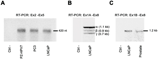
Figure 4. SHBG expression in human prostate cell lines.
A) RT-PCR analysis of SHBG expression in human prostate-derived cell lines, using primers that amplify a region common to all SHBG mRNA isoforms (Ex2-Ex5). B) RT-PCR analysis (Ex1A-Ex8) of exon 1A transcripts in LNCaP cells. The different bands observed correspond to: α (full length transcript), β (skipping of exon 7) and γ (skipping of exons 6 and 7). C) As previously reported [2], full-length TU-1B transcripts are detected by RT-PCR analysis (Ex1B-Ex8) in human prostate tissue and LNCaP cells. Negative controls (Ctrl) are performed with water instead of cDNA.

