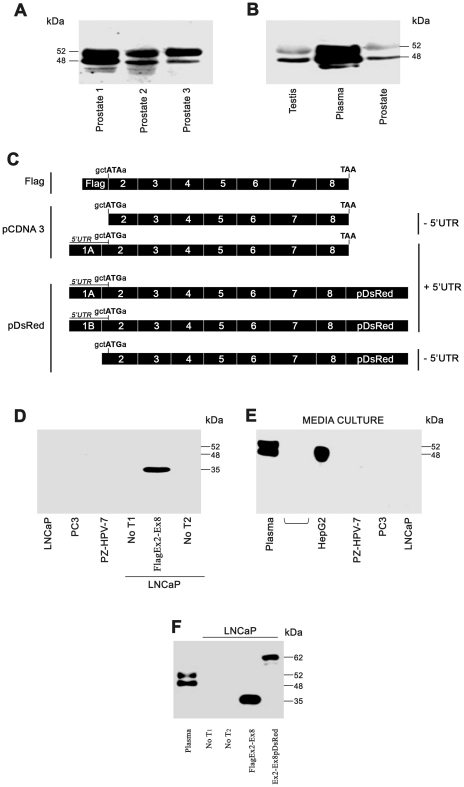
Figure 5. Western blot analysis of SHBG protein in human prostate.
A–B) Two bands sized 52- and 48-kDa were detected in human prostate tissue using the SHBG 11F11 antibody (A) and were identical in size to those detected in human plasma and human testis (B). C) SHBG constructs used to transfect LNCaP cells. In the Flag-Ex2-Ex8 construct, SHBG ATG was mutated to ATA to avoid disturbing the Flag translation start site. The nucleotides involved in the SHBG translation start site are shown in all the constructs. D−E) Western blot analysis of the SHBG protein using the 11F11 antibody in transfected and non-transfected human prostate-derived cell lines (D), and in the supernatant (E). In (E), plasma and the hepatocarcinoma cell line (HepG2) were used as positive controls for secreted SHBG. F) Western blot analysis of LNCaP cells transfected or not with Flag-SHBG and pDsRed-SHBG constructs.

