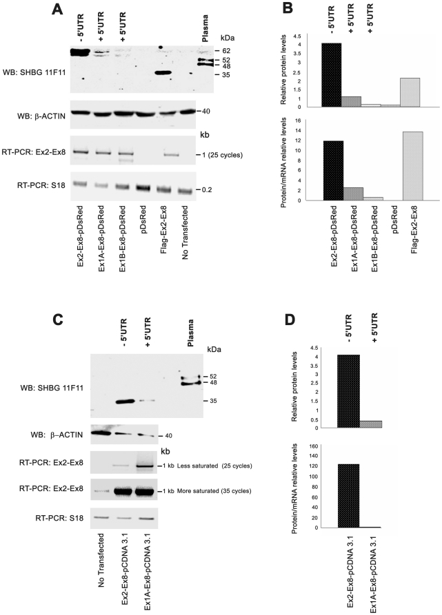
Figure 6. SHBG translation is differentially modulated by alternative exons 1A and 1B.
A) Western blot analysis of SHBG translation in LNCaP cells transfected with different SHBG-pDsRed constructs, with and without different 5′UTRs (exon 1A or exon 1B). Human plasma and Flag-SHBG constructs were used as positive controls of SHBG protein detection, while LNCaP cells transfected with pDsRed empty vector and non-transfected cells were used as negative controls. β-actin was used to normalize the quantity of protein loaded on the acrylamide gel. RT-PCR analysis using primers that recognize a region common to all SHBG isoforms (Ex2-Ex8) was performed to normalize the transfection levels with the different SHBG constructs. S18 primers were used to normalize cDNA levels loaded into the PCR reaction. B) Relative protein levels (RPL) and relative protein/mRNA levels (RPML) are shown. RPL was calculated as a ratio of the β-actin level in the sample. RPML was obtained by dividing RPL by the quantified intensity of mRNA SHBG bands. C) Western blot analysis of SHBG translation in LNCaP cells transfected with pcDNA 3-SHBG constructs with and without the 5′UTR (exon 1A). D) RPL and RPML were calculated as above.

