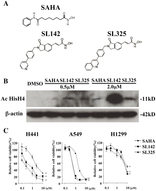
Figure 1. SL142 and SL325 significantly suppressed cell viability in H441 and A549 lung cancer cells.
A. Chemical structure of SAHA, SL142 and SL325. B. Detection of H4 acetylation by immunoblot 24 hours after SAHA, SL142 or SL325 treatment (0.5 or 2.0 µM) in H441 lung cancer cells. β-actin is shown as control. C. Effect on cell viability induced by SAHA, SL142 or SL325. Cells were plated in 96-well plates at a density of 1×10³ cells/well 24 hours prior to treatment with SAHA, SL142 or SL325 (0.1 to 10 µM). Cell viability was evaluated at 96 hours following treatment by the WST1 assay (Roche, Basel, Switzerland) according to the manufacturer's protocol. **, significant difference from the cell viability treated with 0.1 µM of SAHA, SL142 or SL325 (p<0.01).

