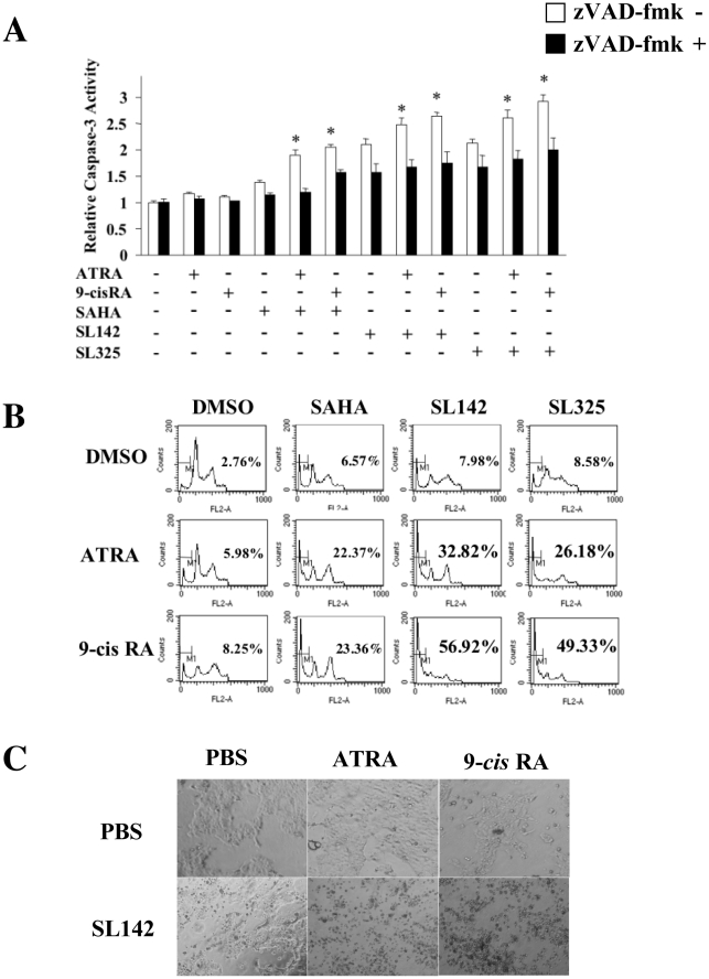
Figure 3. Combination treatment of retinoic acids and SL142 or SL325 more significantly induced cell death in H441 lung cancer cells than those of single use.
A. Analysis of caspase-3 activity induced by ATRA or 9-cis RA (2.5 µM) and/or SAHA, SL142 or SL325 (0.5 µM) in H441 lung cancer cells. Cells were treated with SAHA, SL142 or SL325 at the concentration of 2.5 µM for 36 hours. After 36 hours of treatment, sub-G0/G1 DNA content was measured by propidium iodide staining and flow cytometric analysis. Triplicate experiments were performed; data represent the mean-fold increase ± S.E. *, significant difference from control cells (cells without treatments) (p<0.05). B. Flow cytometric analysis of apoptosis induced by ATRA or 9-cis RA (2.5 µM) and/or SAHA, SL142 or SL325 (0.5 µM) in H441 lung cancer cells. After 96 hours of treatment, sub-G0/G1 DNA content was measured by propidium iodide staining and flow cytometric analysis. C. Morphological analysis of H441 cells after ATRA or 9-cis RA (2.5 µM) and/or SL142 (0.5 µM) treatment.

