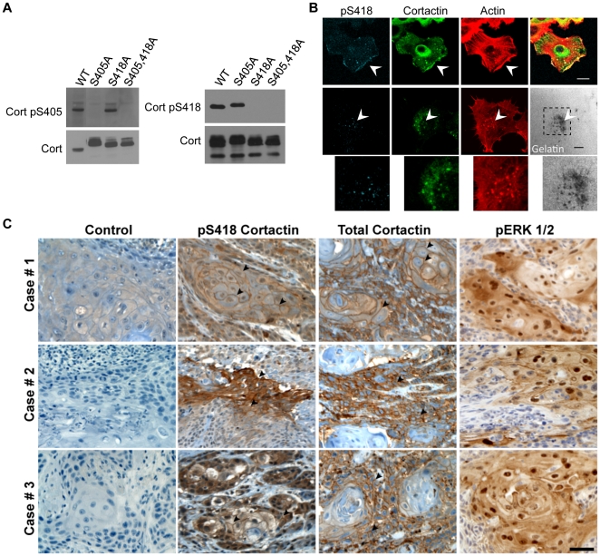
Figure 1. Specificity and validation of pS405 and pS418 phospho-specific cortactin antibodies.
(A) Phospho-specific recognition of anti-cortactin pS405 and pS418 antibodies. Clarified lysates (50 micrograms) from 1483 cells transfected with Myc-tagged wild-type cortactin (WT), Myc-cortactin S405A, Myc-cortactin S418A or Myc-cortactin S405A,S418A point mutants were immunoblotted with affinity purified anti-Cort-pS418 (left) and anti-Cort-pS405 (right) antibodies. (B) Localization of pS418 cortactin in areas of motile and invasive actin dynamics. UMSCC2 cells (top row) were serum starved for 16 h prior to stimulation with 100 nanograms/ml EGF for 1 h to induce lamellipodia formation, while UMSCC1 cells (middle row) were plated on FITC-conjugated gelatin coated coverslips (pseudocolored white) for 6 h to promote invadopodia formation. Cells were fixed, permeablized, and labeled with TRITC-phalloidin (Actin), anti-cortactin (Cort) and anti-cortactin-pS418 antibodies. Arrows denote localization of pS418 cortactin with total cortactin and F-actin in lamellipodia (top) and to invadopodia (middle) coinciding with areas of active matrix degradation. Bottom panels are magnified views of the indicated cellular region. Bars, 10 micrometers. (C) Localization of pS418 cortactin in HNSCC tumor tissue. Serial sections from three different invasive HNSCC cases were processed for immunohistochemistry with control IgG (Control), pS418 cortactin, total cortactin and phospho-ERK1/2 (pERK) antibodies. Sections were counterstained with hematoxylin. Arrowheads indicate areas of peripheral pS418 cortactin and total cortactin enrichment within each tumor sample. Bar, 100 micrometers.

