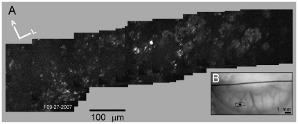
Figure 5. Horizontal scan along medio-lateral axis across the memTNXL injection site (M3R).
(A) Montage of TPSM images with brightly labeled neurons approximately in center. Each YFP image is an average of 10 frames at 512×512 pixels resolution. (B) Location of TPSM scanning sites for montage (small rectangle over medial injection site) on photograph of brain surface.

