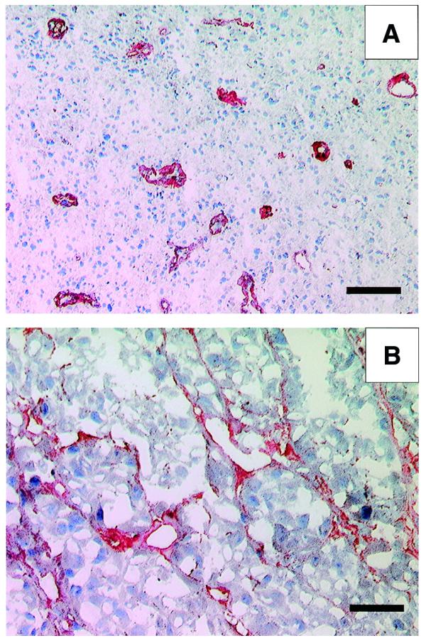
Figure 4.

Immunohistochemical experiments with the antibody ME4C. (A) A section of glioblastoma multiforme specimen. The typical glomerulus-like vascular structures are stained in red by the scFv(ME4C). Scale bar, 100 µm. (B) A section of SKMEL-28 human melanoma, grafted in a nude mouse, stained with scFv(ME4C). The antibody localises around vascular structures and proliferating cells. Scale bar, 50 µm
