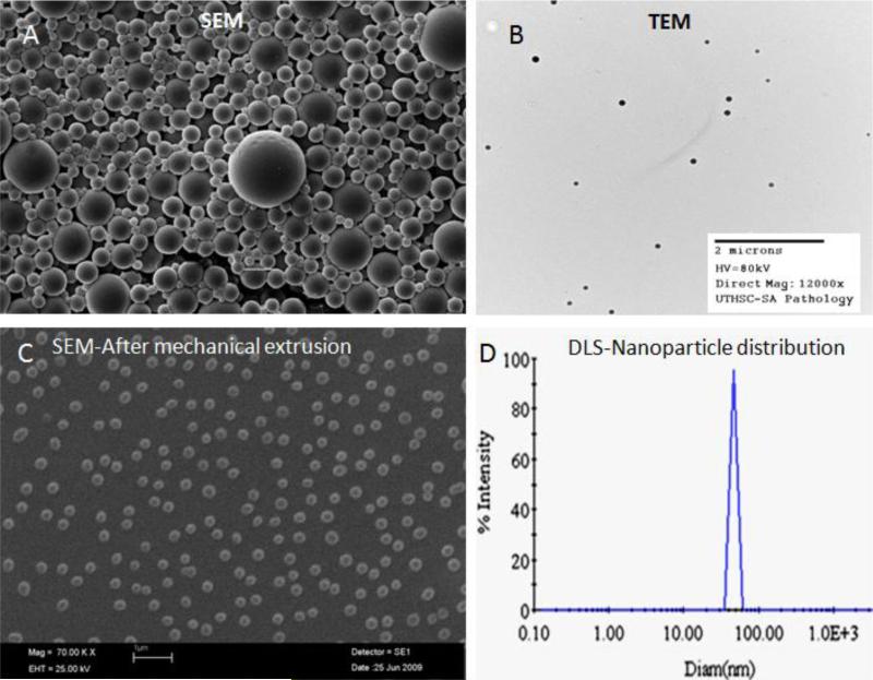
Figure 2. Physical characterization of the nanoparticles.
Some of the commonly used methods to characterize the nanoparticles are depicted in the figure. A- Scanning electron microscopy (SEM) image of nanoparticles; B- Transmission electron microscopy (TEM) image; C- SEM image of nanoparticles after mechanical extrusion; D – Determination of size distribution of nanoparticles by use of dynamic light scattering (DLS).

