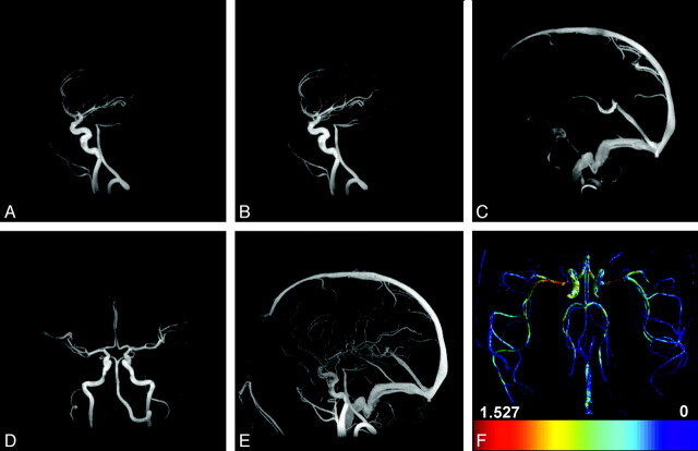
Fig 1.
A, Sagittal HYPRFlow timeframe in the early arterial phase. B, Sagittal HYPRFlow timeframe in the late arterial phase. C, Sagittal HYPRFlow timeframe in the venous phase. D, Coronal HYPRFlow timeframe in the arterial phase. E, Sagittal PC-VIPR MIP. F, WSS map of the axial whole brain generated from PC-VIPR velocity data.

