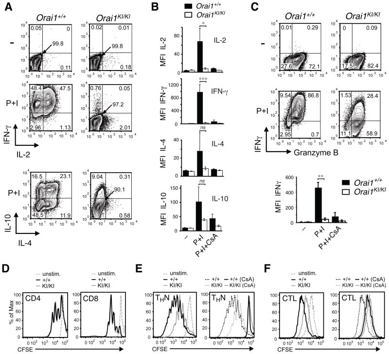
Fig. 7. Impaired effector function of Orai1KI/KI T cells in vitro.
A–C, Severely impaired cytokine production in CD4+ and CD8+ T cells from Orai1KI/KI mice. Expression of the indicated cytokines was measured in CD4+ (A, B) and CD8+ (C) T cells from Orai1KI/KI and Orai1+/+ control mice. T cells were differentiated in vitro under non-polarizing conditions in the presence of 20 U/ml (A, B) and 100 U/ml (C) IL-2. On day 6, cells were left untreated (–) or restimulated for 6 hours with PMA (P) and ionomycin (I) either in the presence or absence of cyclosporin A (CsA). One representative experiment is shown. Bar graphs in B and C represent averages (±SEM) of mean fluorescence intensity (MFI) of cytokine expression from n=4–8 (IL-2, IFN-γ in B), n=3 (IL-4, IL-10 in B) and n=3 (IFN-γ in C) repeat experiments. *, p<0.05; **, p<0.005; ***, p<0.001; ns, not significant. D–F, Normal to moderately impaired proliferation of naïve and in vitro differentiated T cells from Orai1KI/KI mice. D, CD4+ and CD8+ T cells were isolated from lymph nodes of Orai1KI/KI (gray) and Orai1+/+ control (black) mice, loaded with CFSE and stimulated with anti-CD3 and anti-CD28 for 3 days. E–F, CD4+ and CD8+ T cells from Orai1KI/KI (gray) and Orai1+/+ control (black) mice were differentiated into THN cells and cytotoxic T lymphocytes (CTL), respectively, for 3 days in the presence of IL-2, loaded with CFSE and restimulated with anti-CD3 and anti-CD28 for another 3 days without IL-2, either in the presence or absence of cyclosporin A (CsA). Solid lines in right panels of E, F show proliferation of cells in the presence of CsA; proliferation in the absence of CsA (same date as in left panels in E, F) is indicated by dashed lines for comparison. One representative experiment of 3 is shown.

