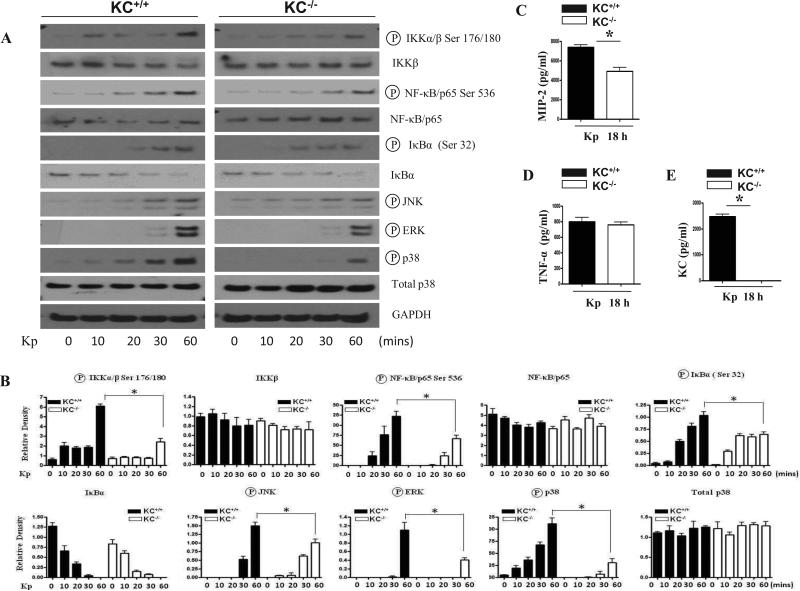
Figure 7. Activation of NF-κB and MAPKs in BMMs of KC-/- mice infected with Kp.
(A) Decreased activation of NF-κB and MAPKs in BMMs obtained from KC-/- mice post Kp infection. Activation of IKK, NF-κB and MAPKs was detected at 24 and 48 h following Kp stimulation. The blot is a representative of three independent experiments with identical results. (B) Densitometric analysis of IKK, NF-κB and MAPK activation in BMMs up to 60 min following stimulation with Kp (MOI of 1). Data expressed as mean ± SE of three blots from three mice in each group (p < 0.05). (C) Attenuated production of MIP-2 in BMMs obtained from KC-/- mice post Kp infection. BMMs were stimulated with Kp at an MOI of 1 and supernatants collected at 18 h post infection were used to determine MIP-2, KC and TNF-α release. The data obtained from 3 independent experiments.

