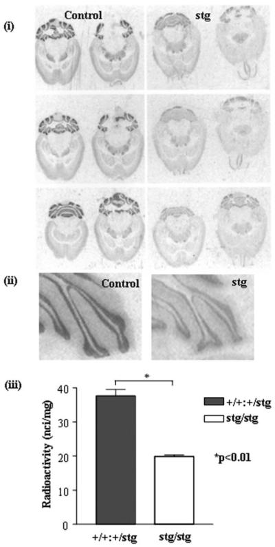
FIGURE 2. [3H]Muscimol binding to control (+/+) and stargazer (stg) adult mouse brain, in situ autoradiography.

i, in situ autoradiography of [3H]muscimol (α6βγ2, α1α6βγ2, α6βδ, and α1α6βδ GABARs) binding in +/+ and stg cerebella using a saturating concentration of [3H]muscimol (20 nm). Nonspecific binding was determined by competitive displacement of muscimol with GABA (100 μm). The signal obtained under these latter conditions was at the level of film background. Six sections from different horizontal planes are shown to demonstrate that the loss of receptor expression was consistent throughout the cerebellum. ii, magnified image of cerebellar lobules labeled with [3H]muscimol. iii, histogram illustrating the results of quantitative image analysis of grayscale intensities using image J software to determine the relative amounts of ligand bound in the cerebellar granule cell layers. A dramatic, significant reduction (by 46 ± 3%, p < 0.01) in [3H]muscimol binding in the cerebellar granule cell layer of stg was determined. Data shown are representative of at least three +/+ and three stg brains and a minimum of 10 sections per brain.
