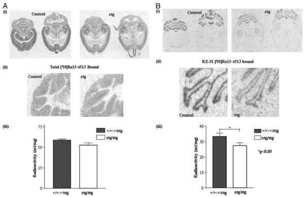
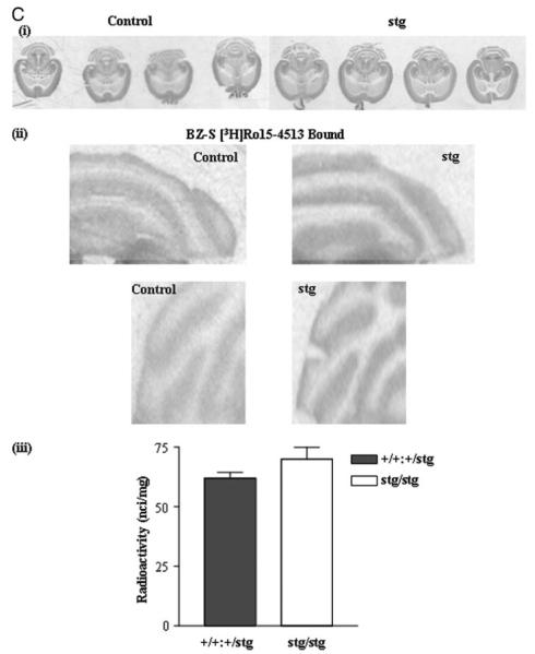
FIGURE 4. [3H] Ro15-4513 binding to control (+/+) and stargazer (stg) adult mouse brain; in situ autoradiography.
A, total [3H]Ro15-4513 binding. Panel i, in situ autoradiography of total [3H]Ro15-4513 binding (γ2-containing GABARs) to +/+ and stg cerebella using a near-saturating concentration of [3H]Ro15-4513 (20 nm). Nonspecific binding was determined by competitive displacement of Ro15-4513 with the benzodiazepine receptor antagonist, Ro15-1788 (10 μm). The signal obtained under these latter conditions was at the level of film background. Two representative comparable sections per mouse strain are shown. Panel ii, magnified image of cerebellar lobules labeled with [3H]Ro15-4513. Panel iii, histogram illustrating the results of quantitative image analysis of grayscale intensities using image J software to determine the relative amounts of ligand bound in the cerebellar granule cell layers. A small (10 ± 4%) but insignificant (p > 0.05) reduction in total [3H]Ro15-4513 binding in the cerebellar granule cell layer of stg was determined. Data shown are representative of at least three +/+ and three stg brains and a minimum of 10 sections per brain. B, BZ-IS [3H]Ro15-4513 binding. Panel i, in situ autoradiography of flunitrazepam (10 μm)-insensitive [3H]Ro15-4513 (BZ-ISR) binding (α6βγ2, α1α6βγ2 GABARs) in +/+ and stg cerebella using a saturating concentration of [3H]Ro15-4513 (20 nm). Nonspecific binding was determined by competitive displacement of Ro15-4513 with benzodiazepine receptor antagonist, Ro15-1788 (10 μm). The signal obtained under these conditions was at the level of film background. Panel ii, magnified image of cerebellar lobules following autoradiography to identify BZ-ISRs. Panel iii, histogram illustrating the results of quantitative image analysis of grayscale intensities using image J software to determine the relative amounts of radioligand bound in the cerebellar granule cell layer. A small but significant reduction (by 21 ± 7%; p < 0.05) in flunitrazepam-insensitive [3H]Ro15-4513 binding in the cerebellar granule cell layer of stg was determined. Data shown are representative of at least three +/+ and three stg brains and a minimum of 10 sections per brain. C, BZ-S [3H]flunitrazepam binding. Panel i, in situ autoradiography of [3H]flunitrazepam (BZ-SR) binding (e.g. α1βγ2 GABARs) in +/+ and stg cerebella using [3H]flunitrazepam (5 nm). Nonspecific binding was determined by competitive displacement of flunitrazepam with benzodiazepine receptor antagonist, Ro15-1788 (10 μm). The signal obtained under these conditions was at the level of film background. Panel ii, magnified image of cerebellar lobules following autoradiography to identify BZ-SRs. Panel iii, histogram illustrating the results of quantitative image analysis of grayscale intensities using image J software to determine the relative amounts of radioligand bound in the cerebellar granule cell layer. A small, nonsignificant increase (by 13 ± 6%; p > 0.05) in [3H]flunitrazepam binding in the cerebellum of stg was determined.


