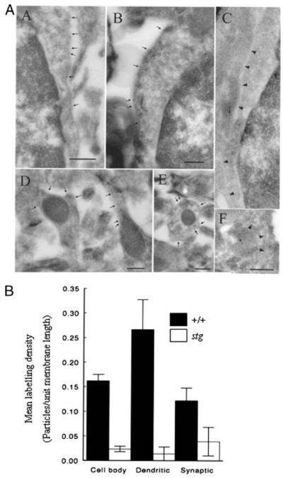
FIGURE 8. Postembedding immunocytochemistry on electron microscopic sections.

A, immunolabeling of GABAR α6 subunit in the cerebellar granule cell layer of +/+ (panels A, B, D, and E) or stg (panels C and F) mouse cerebellum. The plasma membrane of granule cell bodies from +/+ mice (panels A and B) are labeled by gold particles (arrows), but the plasma membrane of stg mice (panel C; arrowheads) are not labeled, and the arrow in panel C marks a gold particle representing nonspecific-labeling of a mitochondrial profile. Plasma membrane of a dendrite (containing mitochondrial profiles) from +/+ mice is strongly labeled but those from stg mice are not labeled (arrow in panel F represents background labeling over a mitochondrial profile). Bars represent 500 nm in A and 250 nm in panels B–F. Two +/+ and two stg brains were fixed by each perfusion method. B, quantitative analysis of mean labeling density (number of gold particles/unit membrane length) of GABAR α6 immunoreactivity in various extrasynaptic (soma and dendrites) and synaptic subdomains of cerebellar granule cells from +/+ (closed boxes) and stg mice (open boxes) in situ. Data are representative of the mean ± S.E.
