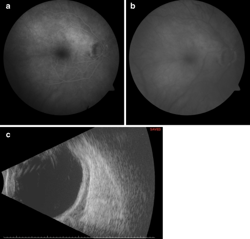
Fig. 1.
a Early-phase fluorescein angiogram of the right macula demonstrating mild hyperfluorescence superotemporal to the optic disk. b Late-phase fluorescein angiogram of the right macula demonstrating diffuse choroidal staining throughout the macula. c B-scan ultrasonograpy of the right eye demonstrating diffuse low-reflective thickening posterior to the sclera, suggesting intraocular involvement

