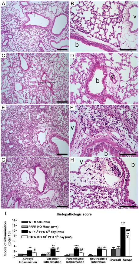
Figure 3. Histological changes after Influenza A WSN/33 H1N1 lethal infection in WT and PAFR deficient mice.
Representative lung slides of WT mock (A, B), PAFR KO mock (C, D), WT (E, F) and PAFR deficient mice (G, H) infected with 106 PFU, after five days of infection. Perialveolar infiltration, vascular and parenchyma inflammation induced by Influenza A virus infection are reduced in PAFR KO mice. Pictures on the left (A, C, E, G) were taken under 100× magnification, bars represent 25µm. Pictures on the right (B, D, F, H) were taken under 400× magnification in the areas highlighted in the lower magnification, bars represent 10µm. Bronchiole and vessels are indicated with “b” and “v”, respectively. Histopathological score (maximal of 18) evaluated airway, vascular, parenchymal inflammation, neutrophilic infiltration (I). Data are presented as Mean ± SEM. ** and *** for p<0.01 and p<0.001, respectively, compared to Mock groups; # for p<0.05 and ## for p<0.01 compared to WT infected group (one-way ANOVA, Newman-Keuls).

