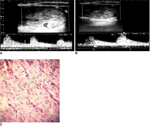
Fig. 4.
Hypoechoic, solid nodule with smooth borders. Although there is no halo or calcification, it was diagnosed as medullary carcinoma. It has mixed vascular pattern in power Doppler US. Pulsatility index and resistive index values calculated by spectral Doppler US were 1.26-0.73 (A) and 1.63-0.80 (B) in center and periphery of nodule, respectively. Isles of cells with large nuclei and wide granular cytoplasm showed marked pleomorphism (C).

