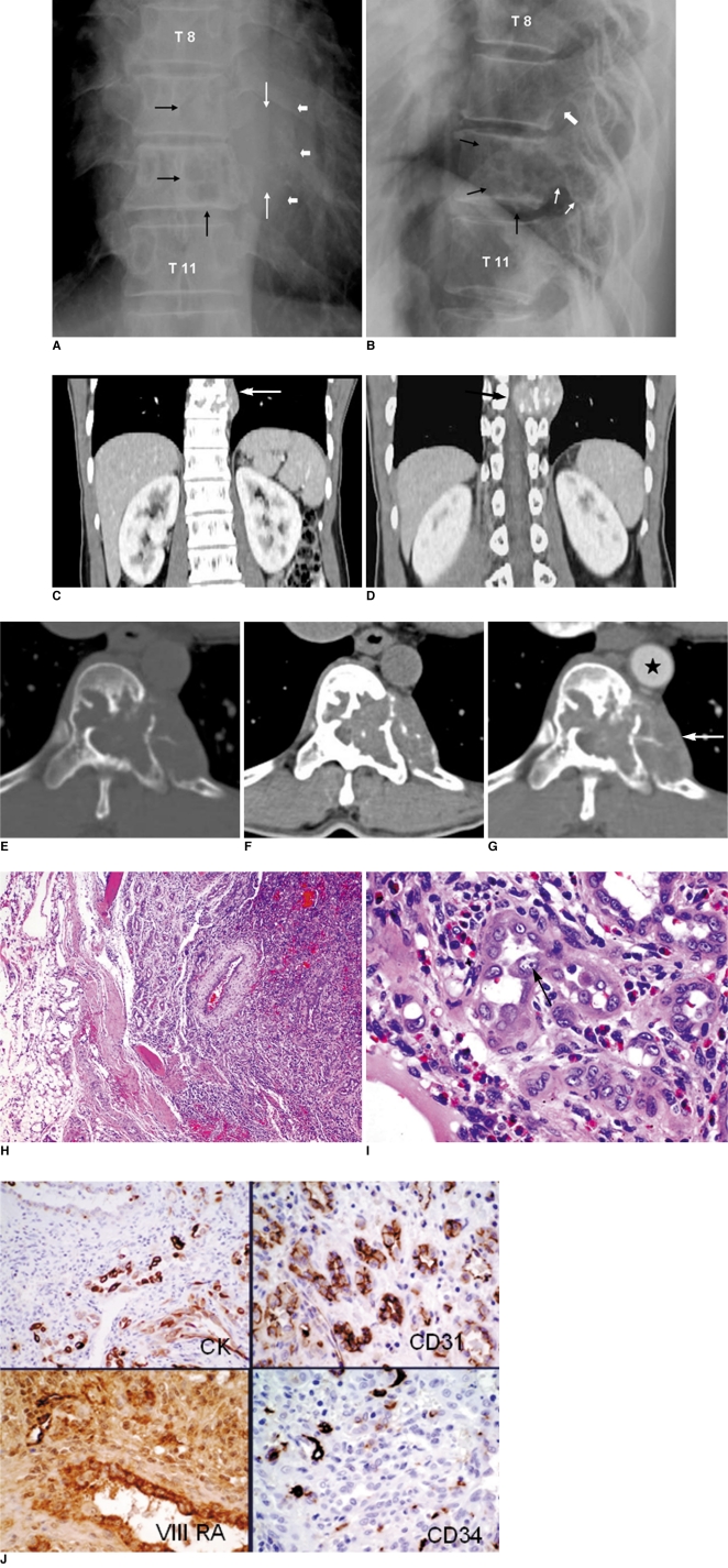
Fig. 1.
Epithelioid hemangioma in 48-year-old man.
Plain radiographs. Anteroposterior (AP) view (A) reveals geographic osteolytic lesion with partially sclerotic border involving left side of T10 vertebral body. Another similar osteolytic lesion is also observed on left side of T9 vertebral body (black arrows). In addition, permeative osteolytic lesion involving posterior part of left 10th rib at costo-vertebral junction (thin white arrows), associated with soft tissue mass (thick white arrows) was observed. No periosteal reaction is observed. Lateral view (B) reveals lesion involving posterior portion of T10 vertebral body (black arrows) with extension to posterior element (thin white arrows). Osteolytic lesion is also seen involving posterior portion of T9 vertebral body (thick white arrow).
C-G. Coronal CT scan (C, D) after iodinated contrast medium injection reveals that lesion involves two contiguous vertebral bodies, T10 and T9 (arrow in C), which causes spinal cord compression (arrow in D). Axial CT scan at T10 vertebral body viewing in bone window (E) and soft tissue window before (F) and after (G) contrast medium injection reveals multiple internal trabeculations, multiple scattered tiny calcifications, and mild enhancement of lesion. Left-side cortex of T10 vertebral body and ventral cortex of 10th rib were destroyed with soft tissue component forming as extrapleural mass (arrow in G) behind thoracic aorta (star).
H. Photomicrograph of lesion under low power (×40) reveals well-circumscribed lobular lesion made up of crowded small tubular or gland-like structure surrounded centrally by large vessel with thick wall. Periphery of lesion is surrounded with fibrofatty tissue.
I. Under ×400 magnification, lining cells of gland-like structure show irregular vesicular or optically clear nuclei with prominent basophilic nucleoli and occasional vacuolated cytoplasm. Tall and enlarged endothelial cell mimicking 'tomb-stone like structure' projected into lumen, is observed (arrow).
J. Immunostains using antibodies reacting to epithelial marker (CK) and vascular markers (CD31, CD34 and Factor VIII RA), show that lining cells are strongly marked by all of these antibodies.

