
Figure 6.
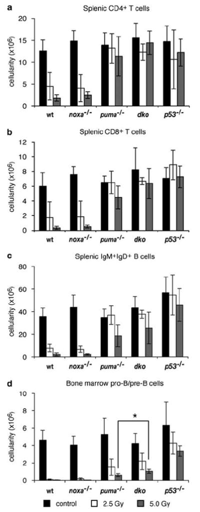
Sensitivity of lymphocytes from noxa−/−puma−/− mice to γ-irradiation in vivo. Wt, noxa−/−, puma−/−, noxa−/−puma−/− or p53−/− mice were left untreated or exposed to 2.5 or 5 Gy full-body γ-irradiation and spleen and bone marrow were harvested 20 h later. (a–c) The number of CD4+ or CD8+ T cells and sIgM+sIgD+ B cells in the spleen from untreated and irradiated mice of each genotype was determined by multiplying the total splenic cellularity with the percentage of the cell subsets. (d) The number of immature B220+sIgM−sIgD− pro-B/pre-B cells in the bone marrow from both femora was calculated using the cellularity and percentage of cell subsets. Bars represent means±S.D. of 4–14 mice of each genotype per treatment in at least three independent experiments. The total number of B220+sIgM−sIgD− pro-B/pre-B cells in the bone marrow of noxa−/−puma−/− animals treated with 5 Gy γ-radiation was significantly greater than in puma−/− animals treated with the same dose (P<0.02)