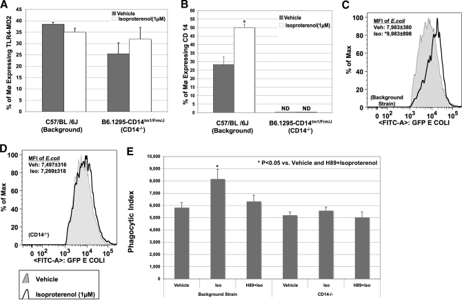
Figure 5. CD14 and not TLR4-MD-2 complex is altered by isoproterenol.
BMØ from CD14−/− mice and background strain stimulated with isoproterenol or vehicle were incubated with ultra-pure LPS or E. coli for 30 min. Staining with F4/80-FITC, TLR4-MD-2-PE-Cy7, and CD14-APC was followed by FACS analysis. Percentage of all F4/80+ BMØ expressing the TLR4-MD-2 complex or membrane CD14 was determined by FACS analyses. (A) Percentage of F480+ BMØ expressing the TLR4-MD-2 complex is not altered by isoproterenol in CD14−/− mice or the background strain. (B) Isoproterenol treatment augments CD14 expression in BMØ obtained from background strain and not CD14−/− mice. Data are given as mean ± sem. *P < 0.05 versus vehicle; n = 4. (C and D) Histograms representing the MFI of phagocytosed GFP-E. coli in F480+ BMØ from the background strain and CD14−/− mice, respectively. Black lines represent isoproterenol treatment, and shaded areas represent vehicle control. *P < 0.05 versus vehicle. (E) Isoproterenol-induced, live E. coli phagocytosis is mediated by PKA signaling. BMØ, obtained from CD14-intact and CD14−/− mice, were pretreated with H-89 (25 μM) before adding isoproterenol (1 μM) or vehicle overnight, and E. coli phagocytosis was determined. The bar graph is the representation of the PI (mean±sem). Blocking with H-89 completely abrogated an isoproterenol-mediated increase in PI in BMØ obtained from CD14-intact background mice, and neither isoproterenol nor H-89 had any effect on CD14−/− mice. *P < 0.05 versus vehicle; n = 4.

