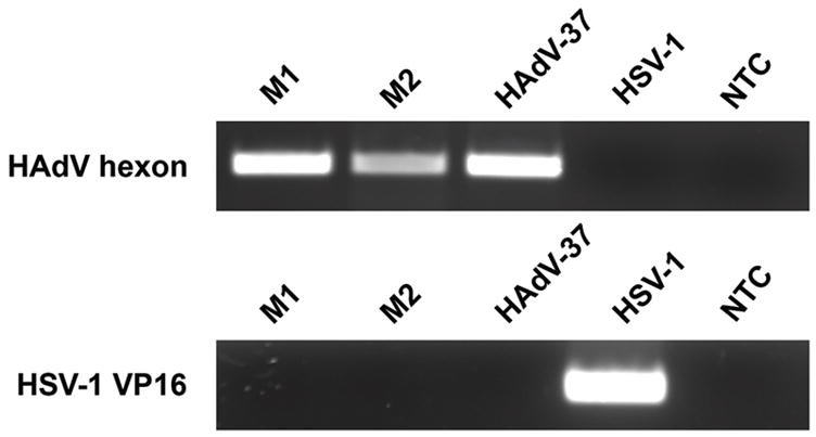
Figure 1.

Identification of HAdV DNA in patient samples by PCR. Samples from both patients (M1 and M2) showed presence of HAdV (top panel). HAdV-37 DNA was used as a positive control, and HSV-1 VP16 gene (bottom panel) as a negative control. Neither membrane contained HSV-1. NTC: no template control.
