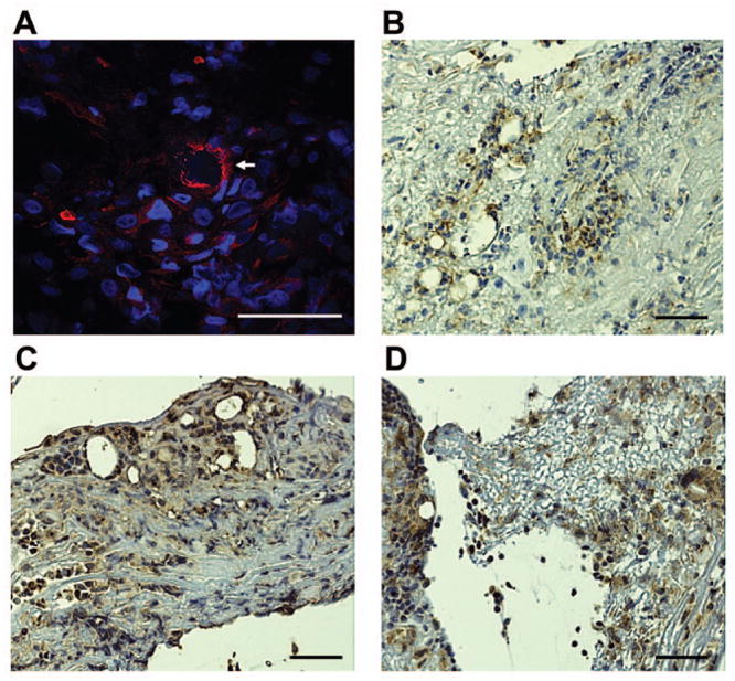
Figure 4.

Angiogenesis in conjunctival membranes in EKC. (A) Representative confocal microscopic image showing Tie-2 staining. Numerous foci of cells positive for Tie-2 are evident, along with a less common circular pattern of staining (arrow). Immunohistochemistry for (B )CD31, (C) TGF-β, and (D) VEGF. Scale bar, 50 μM.
