Figure 5.
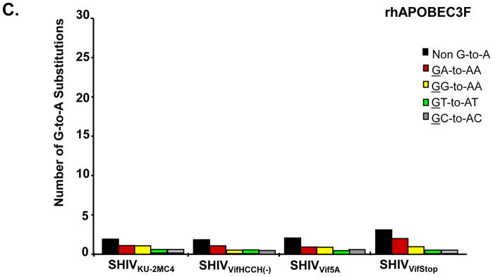
Rhesus A3G but not rhA3F induces G-to-A mutations in single colony sequences obtained from SIVmac239 nef. Full length genomes of the wild type and vif mutant viruses were co-transfected with vectors expressing either rhA3G or rhA3F into 293 cells. At 48 hours post-transfection, the culture medium was collected, clarified by low speed centrifugation and used to infect TZM-bl cells. A 325 base pair fragment of SIVmac239 nef was amplified from DNA at 24 hours post-infection and assessed for G-to-A substitutions. Panel A. G-to-A mutations obtained from bulk (top line) and 15 independent clone sequences are shown. Each mutation is denoted by a vertical line that is color coded with respect to the dinucleotide context: GA (red), GG (yellow), GT (cyan), GC (gray) or non-G-to-A (black). B. Graph depicting the cumulative number of mutations from the 15 clones per virus in the presence of rhA3G. Each bar is shaded according to the proportion of G-to-A-substitutions that occurred in the context of GA (red), GG (yellow), GT (Cyan), GC (gray) or non-G-to-A (black). Panel C. Graph depicting the cumulative number of mutations from the 15 clones per virus in the presence of rhA3F.


