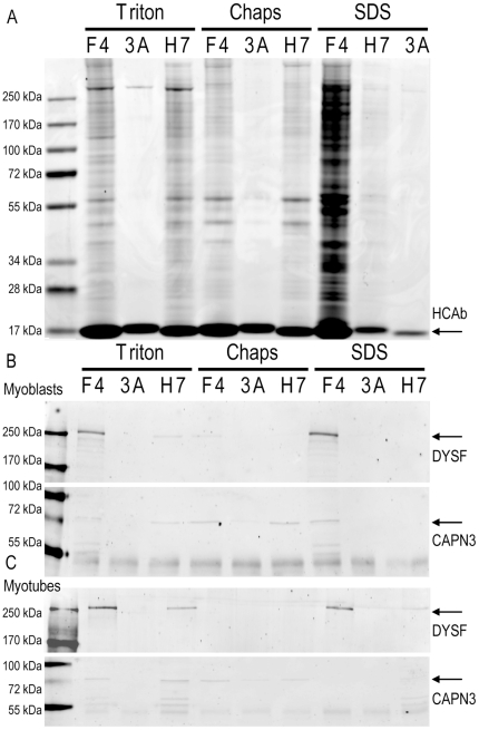
Figure 2. Reproducible dysferlin immunoprecipitation under different conditions.
IM2 myoblasts and myotubes were lysed in three different buffers and subjected to a HCAb Dysferlin immuno precipitation protocol. F4 and H7 are specific for Dysferlin while 3A is a non-specfic control HCAb. A) Coomassie stained gel of immunoprecipitation fractions from myoblasts. B) western blot for Dysferlin and Calpain 3 corresponding to the gel in A. C) A similar western blot on myotubes IP fractions (corresponding gel not shown).

