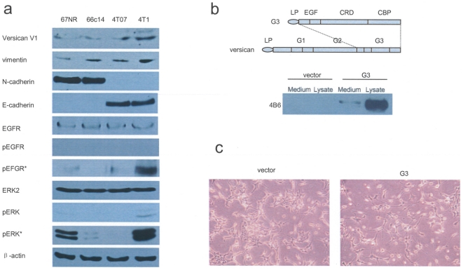
Figure 1. Structure of versican G3 domain and variable versican expression in mouse mammary tumor cells.
(a) Immunoblotting showed that 4T1 cells expressed highest level of versican V1 isoform, vimentin, pERK; 67NR and 66c14 cells expressed N-cadherin, while 4T07 and 4T1 cells expressed E-cadherin; In 20 ng/ml EGF medium, 4T1 cells expressed increased pEGFR and pERK. EGFR*: adding 20 ng/ml EGF for 5 min; ERK*: adding 20 ng/ml EGF for 60 minutes. (b) Versican G3 construct (upper) was expressed in 66c14 cells, analyzed by western blot using cell lysate and culture medium (lower). (c) Morphologically, the G3-transfected 66c14 cells appeared more elongated and spreading more evenly as compared with the predominantly cuboid appearance of cells in the control group that tended to aggregate into clusters.

