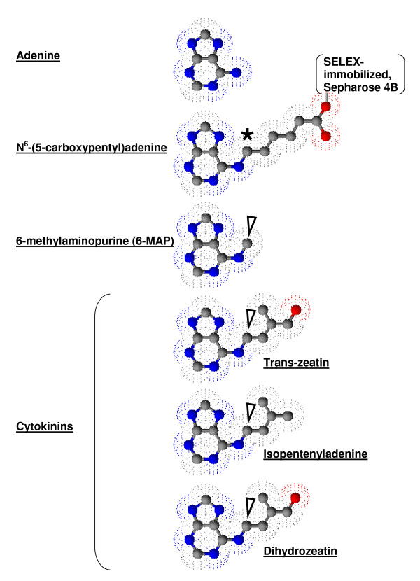
Figure 1.
Structure of Adenine and related compounds, N6-(5-carboxypentyl)adenine, 6-methylaminopurine, and naturally occurring cytokinins. Adenine (top) is compared to the SELEX matrix N6-(5-carboxypentyl)adenine-Sepharose 4B (below). Electron density maps (speckles) indicate the surface RNA could interact with around the ball and stick structures of carbon (grey), nitrogen (blue) and oxygen (red). The black asterisk indicates the N6 substitution anchoring adenine to the SELEX matrix. N6-substituted carbon is also found in the remaining structures (arrows) including 6-methylaminopurine (6-MAP), shown by Meli et al. (2002) to bind N6-(5-carboxypentyl)adenine-Sepharose 4B selected aptamers with higher affinity than adenine. Three major naturally occurring cytokinins are also included.

