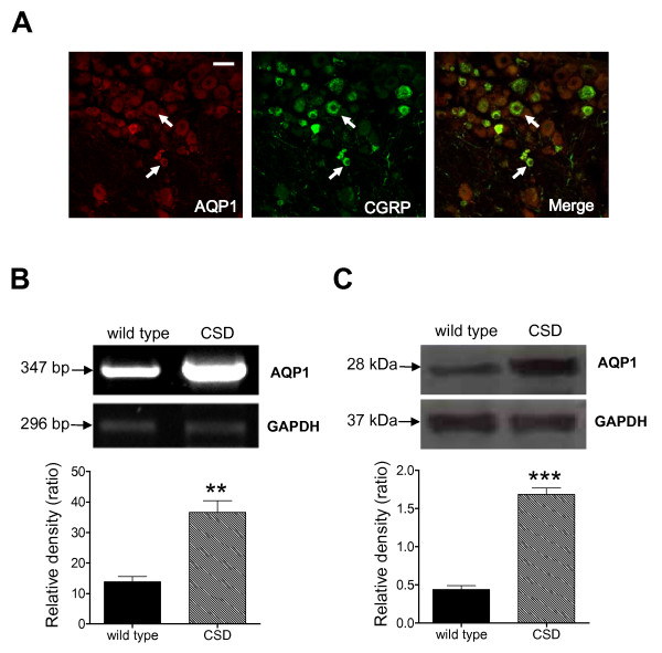
Figure 3.
The expression of AQP-1. A., Co-localization of AQP-1 and CGRP in mouse TG neurons. Merge of double labeling of AQP-1 positive (left, red) and CGRP positive (middle, green) were shown in yellow (right). Bar = 50 μm. B, AQP-1 mRNA was detected in mouse upper cervical and medullary dorsal horn under control and CSD conditions. CSD dramatically upregulated AQP-1 mRNA expression (n = 3, **P < 0.01 vs. wild type). C, AQP-1 protein expression was detection in mouse TNC under control and CSD conditions. CSD greatly enhanced the AQP-1 protein expression in mouse TNC (n = 4, ***P < 0.001 vs. wild type).

