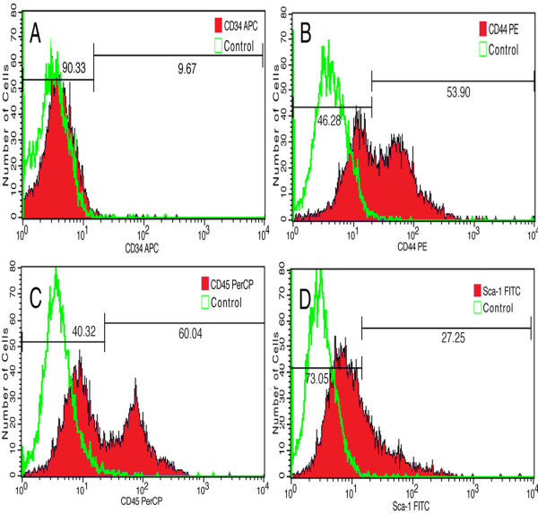
Figure 1.

Fluorescence-activated cell sorting analyses the expression of mesenchymal stem cell markers. APC-conjugated anti-mouse CD34 reactivity is detected on 9.67% of the isolated cells (A). PE-conjugated anti-mouse/human CD44 reactivity is present on 53.90% of the cells (B). Percp-conjugated anti-mouse CD45 antibody labels 60.04% of the cells (C), and FITC-conjugated anti-mouse LY-6A/EC Sca-1 antibody reacts to 27.25% of the cells (D).
