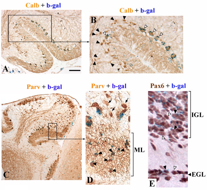
Figure 6. tmgc26<->Rosa26 chimeras suggest cell autonomous effects of the mutation in Purkinje cells and molecular layer interneurons.
(A) Calbindin and β-gal double staining of the cerebellum of chimera X2006 at P19. (B) Higher magnification of the region boxed in panel “A”. Open arrowheads point to β-gal-positive (wild type) Purkinje cells. Black arrowheads point to mutant Purkinje cells. Only wild type Purkinje cells have a normal placement, orientation and size, whereas calbindin-positive β-gal-negative (mutant) cells are small, misoriented and many are displaced in the IGL. (C) Parvalbumin and β-gal double staining of the cerebellum of chimera X2009 at P21. (D) Higher magnification of the region boxed in panel “C”. Virtually all the parvalbumin positive molecular layer interneurons are wild type (β-gal-positive, black arrowheads). The only Parvalbumin-positive/β-gal- negative cells are the displaced Purkinje cells (arrows). (E) Pax6 and β-gal double staining of the cerebellum of chimera X1995 at P19. Both wild type (βgal-positive, black arrowheads) and mutant (βgal-negative, open arrowheads) granule cells were found within both the EGL and IGL. Scale bar: A, 160 μm; B, 70 μm; C, 320 μm; D, 50 μm; E, 40 μm.

